
Core fucosylation (CF) of N-linked glycoproteins is linked with the functions of glycoproteins in many physiological and pathological processes. This feature has a high potential for use in the detection of cancer as well as the development of targeted therapies. For example, high levels of alpha-fetoprotein (AFP)-L3, the AFP with core fucose, allows for the early detection of hepatocellular carcinoma (HCC) with much more specificity than AFP alone, which can also be upregulated in noncancerous liver diseases. However, quantitative characterization of this modification remains challenging due to the complexity and heterogeneity of N-linked glycosylation. In their recent publication, a team of CPTAC researchers from Johns Hopkins University outlines a mass spectroscopy-based method called STAGE that employs sequential treatment of intact glycopeptides with enzymes to analyze site-specific CF of glycoproteins. The team benchmarked their method using samples of both HCC and pancreatic ductal adenocarcinoma (PDAC) which resulted in the identification of over 1900 total CF glycosites.
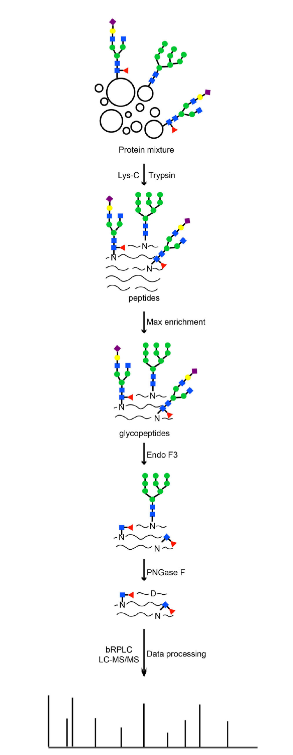
Schematic diagram of the
STAGE method workflow
The STAGE method utilizes Endo F3 followed by PNGase F treatment to generate mass signatures for glycosites that were originally modified by CF N-linked glycans. Using this technique, a total of 1130 unique CF glycosites were identified from human liver tumors and normal tissue. Among the 1130 CF glycosites, 96 CF glycosites were identified (with >1 PNGase F-modified glycosites on the same glycopeptide) which went completely undetected or were detected unreliably in previous studies. Additionally, their proposed method was proven to accurately capture multi-glycosylation sites that occurred simultaniously on the same glycopeptide.
Their quantitative analysis of glycosites from tumor and normal tissues by TMT labeling coupled with the STAGE method allowed the group to document differential expression of glycosylation sites that were up- and down-regulated (FDR < 0.01 and fold change >1.5) in tumor relative to normal tissues. In HCC samples, they observed 88 and 24 CF glycosylation events that were up- and down-regulated in tumor tissues relative to normal, respectively. In PDAC samples they found 54 upregulated and 31 downregulated CF glycosites in PDACs relative to paired normal adjacent tissues. Among these, many were previously identified as having particular significance to their respective disease states such as the glycosite (N603) of EGFR in HCC tumors and protiens in the PI3K-Akt signaling pathway in PDAC tumors. This finding further supports the potential clinical appliaction of this method as a means of detecting and quantifying CF of N-linked glycoproteins. When asked about the significance of the publication, study leader Dr. Hui Zhang proposed “the potential applications of our method span across multiple tumor types, which will be investigated in the future."