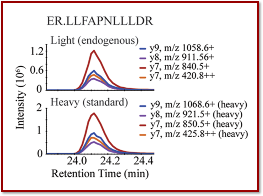
In a recent publication in the journal Clinical Chemistry, CPTAC investigators from Fred Hutchinson Cancer Research Center described a robust technique for evaluating biomarker expression of key receptors in patients with breast cancer bone metastasis using non-decalcified bone biopsies in immuno multiple reaction monitoring mass spectroscopy (immuno-MRM-MS).
Bone is a common site of metastasis in breast cancer. Due to discordance between biomarker expression in primary and metastatic tumors, biopsies of bone metastases are performed prior to treatment to reassess estrogen receptor (ER), progesterone receptor (PR), and human epidermal growth factor receptor 2 (HER2) to guide treatment.
Bone decalcification is a technique used to break down bone tissue for immunohistochemistry (IHC) and fluorescence in situ hybridization (FISH) analysis – two of the most used techniques for evaluating bone metastasis in cancer. However, decalcification of patient bone can also modify or even destroy some proteins, making downstream analysis challenging.
In a blinded study using bone from core needle biopsies of 16 breast cancer patients, CPTAC researchers tested the feasibility of using non-decalcified bone in immuno-MRM-MS for quantitation of estrogen and progesterone receptors, and human epidermal growth factor receptor 2 (HER2) biomarkers. With a mixture of monoclonal antibodies targeting proteotypic receptor peptides prior to immuno-MRM-MS analysis, researchers were able to demonstrate reliable quantification of each biomarker in this proof-of-principle study – even where IHC analysis had failed.
This technique provides a quantitative alternative to IHC and FISH methods in analyzing bone metastasis in breast cancer patients and shows great promise as a clinical approach.

