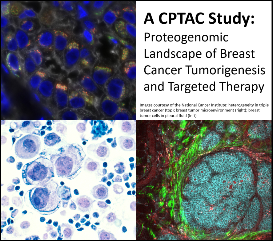
Researchers at Baylor College of Medicine, the Broad Institute of MIT and Harvard and other institutions have applied powerful proteogenomics approaches to better understand the biological complexity of breast cancer. With this approach, the researchers were able to propose more precise diagnostics for known treatment targets, identify new tumor susceptibilities for translation into treatments for aggressive tumors and implicate new mechanisms whereby breast cancers resist treatment. The study appears in the journal Cell.
Proteogenomics combines laboratory techniques  for next-generation DNA and RNA sequencing with mass spectrometry-based analysis for deep, unbiased quantification of proteins and protein modifications in cancer cells, along with computational methods for integrated analysis of this data. Such proteogenomic approaches have been extensively applied to interrogate cancers by investigators at the National Cancer Institute’s Clinical Proteomic Tumor Analysis Consortium (NCI-CPTAC).
“Importantly, our analysis included identification of phosphorylation and acetylation, protein modifications that reveal information about the activity of individual proteins. Protein acetylation had not been profiled in breast cancer before,” said co-corresponding author Dr. Matthew Ellis, breast cancer oncologist and professor and director of Baylor College of Medicine’s Lester and Sue Smith Breast Center, McNair Scholar at Baylor and Susan G. Komen Scholar.
Simultaneously analyzing changes in the genetic code and the manifestations of these cancer-specific alterations in terms of protein function provides a much more complete picture of what is going on inside breast cancer tumors than analyzing each component in isolation.
The researchers’ initial proteogenomic analysis of breast cancer using residual samples from the Cancer Genome Atlas (TCGA) provided proof-of-principle that proteogenomics represented an advance in breast cancer profiling. The current study represents a major step forward in that it included tissue samples that were collected using protocols that specifically preserve protein modifications, analyzed many more samples, carried out genomics and proteomic characterization on exactly the same tissue fragments and added protein acetylation profiling to protein phosphorylation, DNA and RNA measurements. Proteogenomic analytical techniques have matured substantially in recent years, and those cutting-edge approaches were applied to this dataset.
The researchers completed proteogenomic analyses of 122 treatment-naïve primary breast cancer samples. Their measurements generated a tremendous amount of data ‒ about 38,000 phosphorylation sites and almost 10,000 acetylation sites per tumor ‒ and the researchers used advanced computational methods to analyze and integrate the information. “Complex analyses like these are now routinely being performed on large-scale proteogenomic data sets and we are developing tools to automate the process,” said Dr. D. R. Mani, a co-corresponding author and principal computational scientist at the Broad Institute.
“We describe here proteogenomic characterization of the largest set to date of breast cancer samples that were purposefully collected for these types of analyses, maximizing the fidelity and accuracy of the results,” Ellis said. “Each tumor cell has literally hundreds of genomic changes. Mostly we don’t understand their significance either clinically or biologically. The approach we illustrate enables a deeper and more complete understanding of each individual’s breast cancer.”
For example, the analyses revealed that some subtypes of breast cancer have certain targetable enzymes called kinases that are more heavily phosphorylated than in other cancers, suggesting greater activity and therefore targetability. These analyses included recently identified drug targets such as CDK4/6 and its regulatory contact and the programmed cell death receptors and ligands that are the targets of new immunotherapy drugs. Here analyses identified new sets of estrogen receptor-positive breast cancers that could be treated with these agents. This is significant because currently these agents are restricted to estrogen receptor-negative disease.
Additional analyses raised entirely new insights into the metabolic vulnerabilities of ER+ and ER- breast cancer. “Our global analysis of the acetylproteome, the first in breast tumors, exposed new details of breast cancer subtype-specific metabolism,” said co-corresponding author Dr. Steven A. Carr, Director of Proteomics at the Broad Institute of MIT and Harvard.
The researchers hope that their findings will motivate breast cancer scientists to explore the therapeutic or diagnostic potential of the new biological alterations they have identified in this study. They also are optimistic that their findings will encourage an effort to translate proteogenomics into a cancer-profiling approach that can be used routinely in the clinic to improve diagnosis and treatment. “We believe that proteogenomics approaches will continue to help us to identify new candidate therapeutic targets, better understand the immune landscape of breast and other cancers, gain insights into response and resistance, and ultimate progress towards our goal of personalized cancer care,” noted co-corresponding author Dr. Michael Gillette, a pulmonary and critical care physician at Massachusetts General Hospital and Senior Group Leader in Proteomics at the Broad Institute. “The science is powerful and exciting, but in the end it is what we can deliver to the patient that makes it important.”
Find the complete list of all the contributors to this work and their affiliations, as well as all the financial support for this study in the journal Cell.

